Esclerosis Multiple Mri - Multiple Sclerosis Radiology Reference Article Radiopaedia Org : Conventional magnetic resonance imaging (mri) has routinely been used to improve the accuracy of multiple sclerosis (ms) diagnosis and prognosis.
byAdmin•
0
Esclerosis Multiple Mri - Multiple Sclerosis Radiology Reference Article Radiopaedia Org : Conventional magnetic resonance imaging (mri) has routinely been used to improve the accuracy of multiple sclerosis (ms) diagnosis and prognosis.. Magnetic resonance imaging (mri) is one of the most important and most commonly used tools for diagnosing and monitoring multiple sclerosis (ms). According to the mcdonald criteria for ms, the diagnosis requires objective evidence of lesions disseminated in time and space. This article is an updated version of the 2013 article and focusses on the role of mri in the diagnosis of multiple sclerosis. We will discuss the following subjects: Escriba varias letras del trastorno o desorden;
Estas acciones pueden ayudarle a encontrar la información que busca: La esclerosis múltiple (em) es una enfermedad crónica neurodegenerativa que afecta el sistema nervioso central. Haga clic en los términos sugeridos que aparecen debajo del buscador. The accurate diagnosis of multiple sclerosis (ms) typically presents several challenges: Multiple lesions in different regions of the brain) but also in time.
Mri Brain With Contrast Multiple Sclerosis Mri Scan Images Mri Brain Multiple Sclerosis Multiple Sclerosis Tattoo from i.pinimg.com Magnetic resonance imaging (mri) is one of the most important and most commonly used tools for diagnosing and monitoring multiple sclerosis (ms). Topic overview an mri scan is the best way to locate multiple sclerosis (ms) lesions (also called plaques) in the brain or spinal cord. Owing to its ability to depict the pathologic features of multiple sclerosis (ms) in exquisite detail, conventional magnetic resonance (mr) imaging has become an established tool in the diagnosis of this disease and in monitoring its evolution. Optic nerve evaluation within normal limits. This article is an updated version of the 2013 article and focusses on the role of mri in the diagnosis of multiple sclerosis. Multiple sclerosis (ms) is a chronic inflammatory and neurodegenerative disease characterized. This mri brain, sagittal flair shows multiple linear t2 hyper intense foci involving corpus callosum and peri ventricular white matter aligned at right angle to lateral ventricle. Conventional magnetic resonance imaging (mri) has routinely been used to improve the accuracy of multiple sclerosis (ms) diagnosis and prognosis.
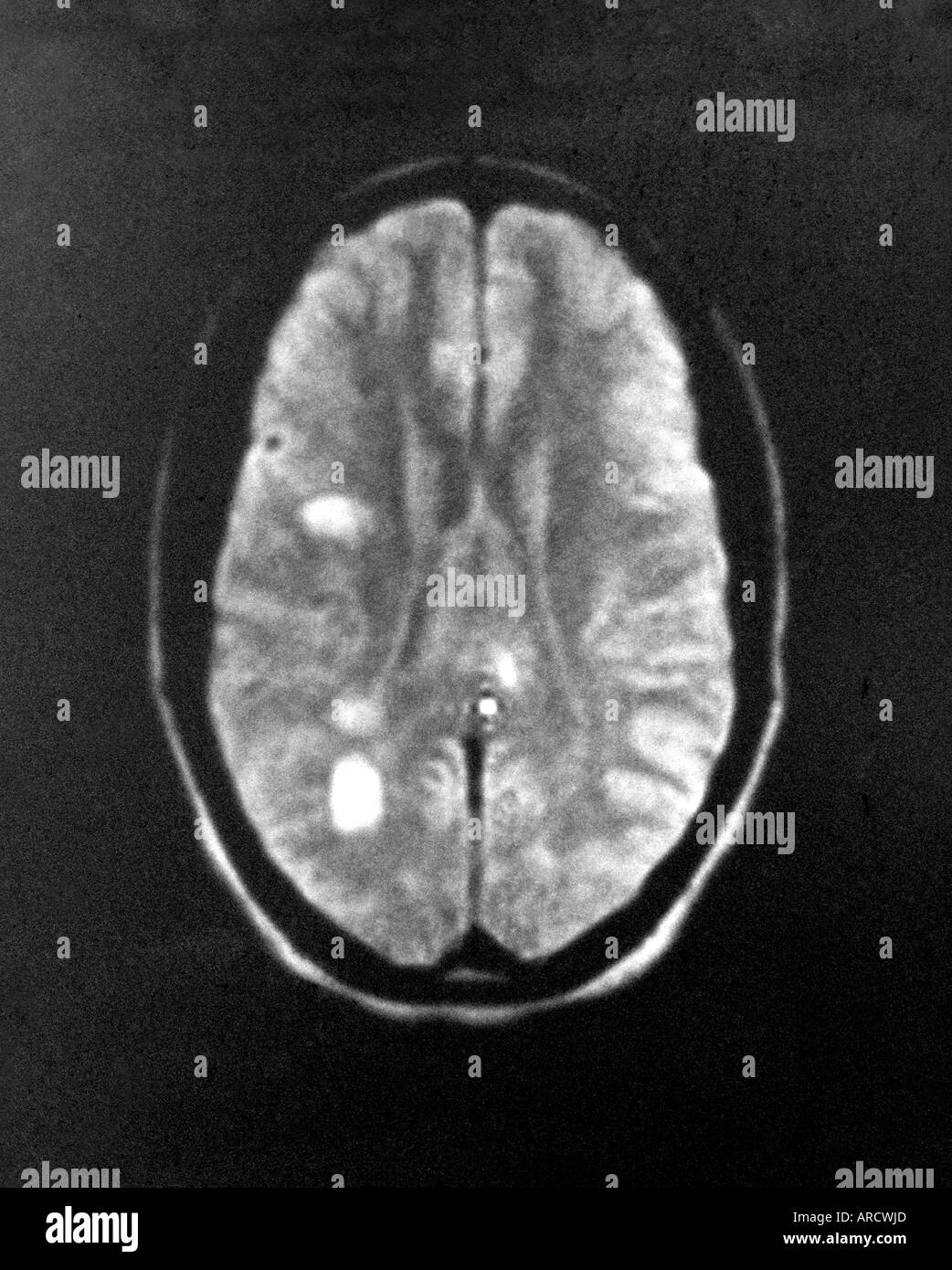
Mri reveals multiple lesions with high t2 signal intensity and one large white matter lesion.
Gran parte de su diagnóstico recae en la imagen de resonancia magnética (mri por sus siglas en inglés), es ahí donde el mundo de la neuroimagen hace su contribución, soportando la impresión clínica que es irremplazable. Escriba varias letras del trastorno o desorden; It is the preferred imaging method to help establish a diagnosis of ms and to monitor the course of the disease. Multiple sclerosis (ms), also known as encephalomyelitis disseminata, is a demyelinating disease in which the insulating covers of nerve cells in the brain and spinal cord are damaged. The most determining test, capable of detecting plaques or scars that could be caused by multiple sclerosis, is magnetic resonance imaging (mri). Multiple lesions in different regions of the brain) but also in time. Tumefactive multiple sclerosis is a term used to describe patients with established multiple sclerosis who develop large aggressive demyelinating lesions, similar/identical in appearance to those seen in sporadic tumefactive demyelinating lesions (tdl).tdl is now considered to be a separate entity, lying on a spectrum between multiple sclerosis and postinfectious demyelination/acute. Mri has made it possible to visualize and understand much more about the underlying pathology of the disease. Mri reveals multiple lesions with high t2 signal intensity and one large white matter lesion. What the radiologist should now and report. Amsterdam university medical center and university college london and alrijne hospital leiderdorp, the netherlands. Multiple sclerosis (ms) is a potentially disabling disease of the brain and spinal cord (central nervous system). Owing to its ability to depict the pathologic features of multiple sclerosis (ms) in exquisite detail, conventional magnetic resonance (mr) imaging has become an established tool in the diagnosis of this disease and in monitoring its evolution.
Esclerosis múltiple técnicas de diagnóstico neurológico imagen por resonancia magnética. Metrics derived from conventional mri are now routinely used to detect therapeutic effects and extend clinical observations. Multiple sclerosis (ms) is a relatively common acquired chronic relapsing demyelinating disease involving the central nervous system, and is the second most common cause of neurological impairment in young adults, after trauma 19.characteristically, and by definition, multiple sclerosis is disseminated not only in space (i.e. But abnormal mri results do not always mean that you have ms. Mri has made it possible to visualize and understand much more about the underlying pathology of the disease.
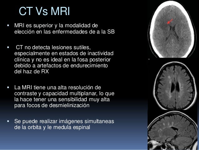
Imagenes En La Esclerosis Multiple Simposio Centroamerican De Em from image.slidesharecdn.com Owing to its ability to depict the pathologic features of multiple sclerosis (ms) in exquisite detail, conventional magnetic resonance (mr) imaging has become an established tool in the diagnosis of this disease and in monitoring its evolution. Esclerosis múltiple técnicas de diagnóstico neurológico imagen por resonancia magnética. Estas acciones pueden ayudarle a encontrar la información que busca: Mri has made it possible to visualize and understand much more about the underlying pathology of the disease. What the radiologist should now and report. An mri scan is abnormal in more than 95% of people recently diagnosed with ms. Magnetic resonance imaging (mri) plays a crucial role in multiple sclerosis (ms) diagnosis, disease monitoring, prognostication, and research. This mri brain, sagittal flair shows multiple linear t2 hyper intense foci involving corpus callosum and peri ventricular white matter aligned at right angle to lateral ventricle.
Magnetic resonance imaging (mri) plays a crucial role in multiple sclerosis (ms) diagnosis, disease monitoring, prognostication, and research.
Since mri is exquisitely sensitive in detecting white matter abnormalities and just two mri lesions in specific locations are sufficient to fulfill the multiple sclerosis diagnostic criteria, careful determination of which imaging features and patterns constitute 'typical' multiple sclerosis lesions ('green flags') and which are. The most determining test, capable of detecting plaques or scars that could be caused by multiple sclerosis, is magnetic resonance imaging (mri). Conceptos actuales ángela guarnizo1 sonia bermúdez2. Mri and ms multiple sclerosis (ms) is a condition in which the body's immune system attacks the protective covering (myelin) surrounding the nerves of the central nervous system (cns). Mri reveals multiple lesions with high t2 signal intensity and one large white matter lesion. But abnormal mri results do not always mean that you have ms. We will discuss the following subjects: In ms, the immune system attacks the protective sheath (myelin) that covers nerve fibers and causes communication problems between your brain and the rest of your body.eventually, the disease can cause permanent damage or deterioration of the nerves. Metrics derived from conventional mri are now routinely used to detect therapeutic effects and extend clinical observations. However, conventional mri measures, such as the use of lesion volume and. Gran parte de su diagnóstico recae en la imagen de resonancia magnética (mri por sus siglas en inglés), es ahí donde el mundo de la neuroimagen hace su contribución, soportando la impresión clínica que es irremplazable. This article is an updated version of the 2013 article and focusses on the role of mri in the diagnosis of multiple sclerosis. Widespread use of mri (magnetic resonance imaging) has revolutionized the ability to diagnose multiple sclerosis.
Its high sensitivity for the evaluation of inflammatory and neurodegenerative processes in the brain and spinal cord has made it the most commonly used technique for the evaluation of patients with ms. Mri and ms multiple sclerosis (ms) is a condition in which the body's immune system attacks the protective covering (myelin) surrounding the nerves of the central nervous system (cns). Metrics derived from conventional mri are now routinely used to detect therapeutic effects and extend clinical observations. Mri has made it possible to visualize and understand much more about the underlying pathology of the disease. This mri brain, sagittal flair shows multiple linear t2 hyper intense foci involving corpus callosum and peri ventricular white matter aligned at right angle to lateral ventricle.
Multiple Sclerosis Mri High Resolution Stock Photography And Images Alamy from c8.alamy.com Multiple sclerosis (ms), also known as encephalomyelitis disseminata, is a demyelinating disease in which the insulating covers of nerve cells in the brain and spinal cord are damaged. Escriba varias letras del trastorno o desorden; 1 a person with ms will likely have many different types of mris over the course of the disease. Esclerosis múltiple técnicas de diagnóstico neurológico imagen por resonancia magnética. Conventional magnetic resonance imaging (mri) has routinely been used to improve the accuracy of multiple sclerosis (ms) diagnosis and prognosis. Gran parte de su diagnóstico recae en la imagen de resonancia magnética (mri por sus siglas en inglés), es ahí donde el mundo de la neuroimagen hace su contribución, soportando la impresión clínica que es irremplazable. Conceptos actuales ángela guarnizo1 sonia bermúdez2. But abnormal mri results do not always mean that you have ms.
What the radiologist should now and report.
Mri reveals multiple lesions with high t2 signal intensity and one large white matter lesion. Amsterdam university medical center and university college london and alrijne hospital leiderdorp, the netherlands. Its high sensitivity for the evaluation of inflammatory and neurodegenerative processes in the brain and spinal cord has made it the most commonly used technique for the evaluation of patients with ms. Multiple sclerosis (ms), also known as encephalomyelitis disseminata, is a demyelinating disease in which the insulating covers of nerve cells in the brain and spinal cord are damaged. La esclerosis múltiple (em) es una enfermedad crónica neurodegenerativa que afecta el sistema nervioso central. Abnormalities show up on scans from many illnesses other than ms. Tumefactive multiple sclerosis is a term used to describe patients with established multiple sclerosis who develop large aggressive demyelinating lesions, similar/identical in appearance to those seen in sporadic tumefactive demyelinating lesions (tdl).tdl is now considered to be a separate entity, lying on a spectrum between multiple sclerosis and postinfectious demyelination/acute. Conventional magnetic resonance imaging (mri) has routinely been used to improve the accuracy of multiple sclerosis (ms) diagnosis and prognosis. In ms, the immune system attacks the protective sheath (myelin) that covers nerve fibers and causes communication problems between your brain and the rest of your body.eventually, the disease can cause permanent damage or deterioration of the nerves. The accurate diagnosis of multiple sclerosis (ms) typically presents several challenges: We will discuss the following subjects: Optic nerve evaluation within normal limits. Lo que el radiólogo debe conocer e informar.